Read carefully.
Follow the sources.
Change the conclusion when the evidence changes.
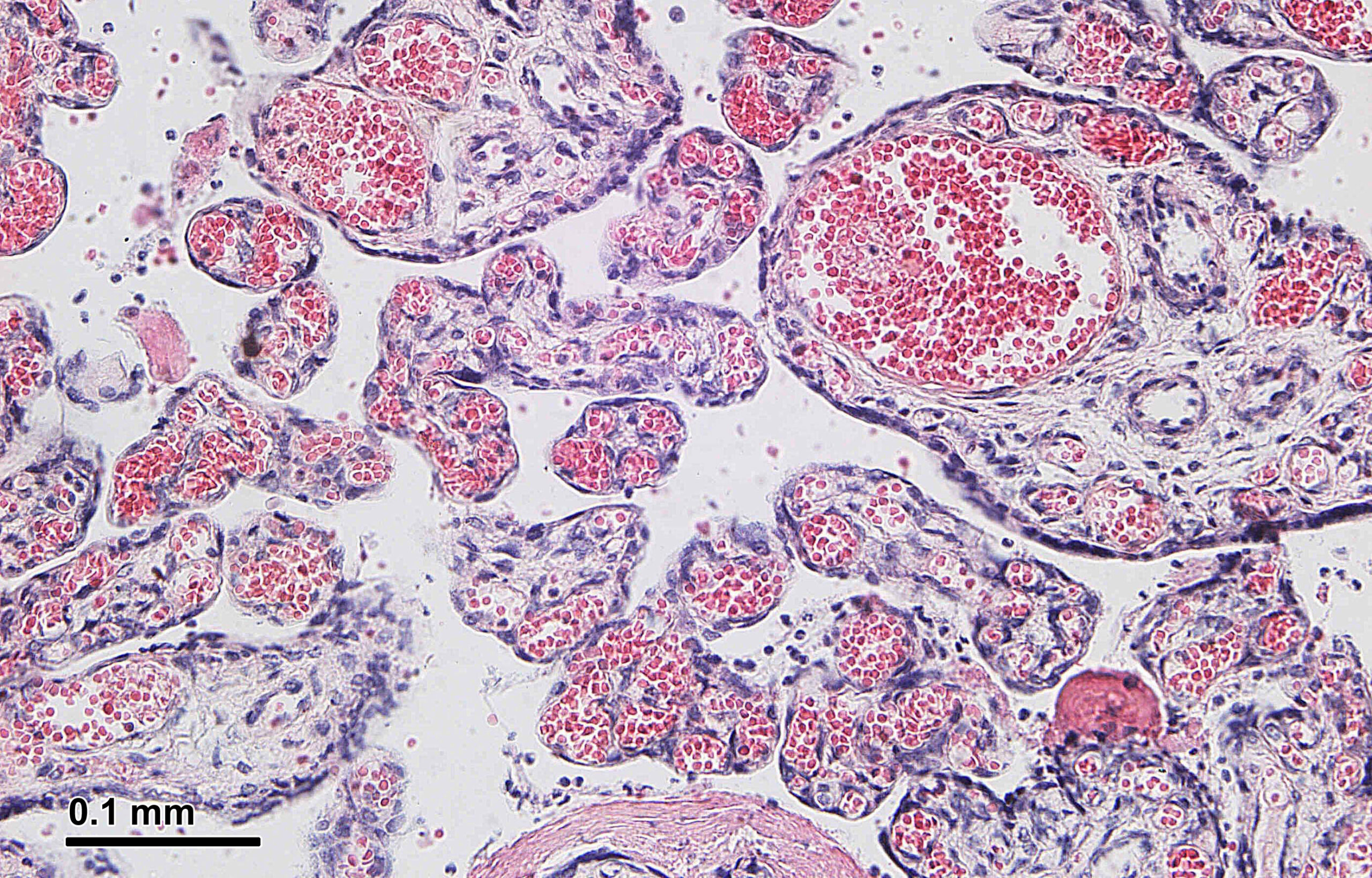
Image: Human placenta, H&E-stained cross-section. Josef Reischig / Wikimedia Commons · CC BY-SA 3.0.
A pregnancy can end while a few of its cells do not. Years later—sometimes decades later—cells carrying genetic markers from that pregnancy may still be detectable in blood or tissue.
That is not a metaphor, and it is not the same as the fragments of cell-free fetal DNA used in prenatal screening. It is a real biological phenomenon called fetal microchimerism: a very small population of fetal-origin cells inside the person who carried the pregnancy. Traffic also runs the other way. Maternal cells can enter the developing fetus and persist into the offspring’s adult life.
So the viral version—“your baby’s cells stay inside you”—has a solid fact underneath it. It also makes the science sound much more settled than it is.
Evidence status
Cells cross the placenta in both directions during pregnancy, and some fetal-origin and maternal cells can persist for decades.
Immune tolerance, cell type and tissue niches probably influence which rare cells survive. Their effects may vary by tissue and circumstance.
Which pregnancies contribute the cells found years later, whether later pregnancies replace earlier populations in humans, and when the cells help, harm or simply linger.
First: the placenta is not a communal swimming pool
Maternal and fetal blood normally remain in separate circulatory systems. They come into extremely close contact at the placenta, where gases, nutrients, waste products, antibodies and other material can cross an interface. A small number of intact cells can cross too.
That distinction matters because “shared bloodstream” suggests indiscriminate mixing. Microchimerism is rarer and more selective. A 2025 group of specialists described microchimeric cells as often occurring at roughly one in a million host cells. Finding them is less like noticing a new ingredient in soup and more like trying to identify one particular person in a city—without always knowing exactly what they look like.
The classic human evidence arrived in 1996. Researchers sorted blood cells from eight women who had given birth to sons between six months and 27 years earlier, then searched for Y-chromosome sequences. They detected male DNA in a progenitor-cell fraction in six of the eight, including the woman whose last son had been born 27 years before. The sample was small and the marker worked only for male-origin cells, but the result established something remarkable: pregnancy can leave a long-lived cellular population, not merely short-lived DNA debris.
Finding a genetic marker is not the same as catching a cell at work
Microchimerism research has a built-in measurement problem. For decades, the easiest foreign marker to spot was a Y chromosome in a female sample—or maternal XX cells in a male sample. That made many studies practical. It also made the field disproportionately good at seeing pregnancies involving male fetuses.
Newer tests can target genetic differences unrelated to sex, including HLA variants and other inherited markers. But each method answers a slightly different question. PCR can tell researchers that a particular stretch of DNA is present and, in quantitative forms, roughly how much. It cannot by itself show that the DNA belongs to a viable, intact cell, reveal what that cell is doing, or place it within a tissue. Imaging can supply location but may be less sensitive and can produce artefacts. Single-cell and spatial methods promise a fuller identity card, but looking for one cell among a million makes them expensive and technically awkward.
This creates an important evidence boundary. “Fetal-origin DNA was detected here” can be strong evidence of microchimerism under a well-designed assay. It is not automatically evidence that a living fetal cell repaired the tissue, attacked it, caused a disease or even came from the pregnancy researchers assume.
Whose cell is it, exactly?
A Y-chromosome marker in someone who once carried a male fetus has an obvious possible source. “Obvious” is not the same as uniquely proven.
Microchimerism can also follow a vanished twin, organ or stem-cell transplantation and, in some circumstances, transfusion. Male-origin cells have been reported in women and girls with no known male pregnancy. Researchers have proposed several routes, including an unrecognised pregnancy, a twin or cells that travelled through the maternal line. The last possibility is especially tempting: perhaps a pregnant person carries cells from her own mother, and some later cross into her fetus.
That multigenerational idea is biologically plausible and supported in some animal models. Direct evidence for routine grandparent-to-grandchild or sibling-to-sibling transfer in humans is still limited. It belongs in the “excellent question” column, not the “secret family cellular network confirmed” column.
Are the cells repairing you—or making you ill?
Researchers have found fetal-origin cells in injured tissue, tumours and organs affected by autoimmune disease. Some experimental work, particularly in animals, suggests certain fetal-origin cells can participate in inflammation, blood-vessel formation and wound repair. Other studies report associations between microchimerism and disorders including systemic sclerosis, thyroid disease or pregnancy complications.
That sounds like two incompatible stories: helpful repair crew versus foreign-cell troublemakers. The evidence supports neither as a universal role.
Diseased and injured tissues send chemical signals that recruit circulating cells. If microchimeric cells arrive because tissue is inflamed, their presence could be a response rather than a cause. A damaged placenta may also allow more fetal cells into maternal circulation, making high cell counts a marker of placental dysfunction rather than the thing producing it. And if a rare cell adopts the appearance of a local cell, that still does not tell us whether it made a meaningful contribution to repair.
The most defensible answer is contextual and mildly unsatisfying: microchimeric cells may be active in some tissues and circumstances, harmful in others, and inconsequential passengers in many. The studies are heterogeneous, cell numbers are tiny, and methods have not been standardised across laboratories. Presence is established. A single health meaning is not.
The neat immune explanation did not stay neat
Long-term survival creates an immunological puzzle. These cells are genetically distinct, yet some escape the rejection that makes organ transplantation so difficult. Regulatory immune cells and compatibility at human leukocyte antigen genes are reasonable suspects.
If HLA compatibility is doing much of the work, certain maternal–fetal HLA combinations should predict which fetal-origin cells remain. A 2025 human study tested variants of that idea. Its headline result was not a clean confirmation: HLA-C groups did not predict microchimerism during or after pregnancy, while one type of HLA-C mismatch was negatively correlated with postpartum cell levels.
That does not rule out immune selection. HLA is a large, complicated system, and one study cannot test every relevant compatibility relationship. It does rule out the most convenient version in which a simple HLA-C match explains who gets to stay.
What if the body does not keep every pregnancy’s cells?
The sentimental picture of microchimerism is an ever-growing cellular scrapbook: each pregnancy adds a few cells, and the collection remains. Mouse research has produced a stranger possibility.
In a 2023 experiment, researchers tracked genetically identifiable cell populations across pregnancies. Maternal-origin cells that female mice had acquired from their own mothers were displaced when the mice became pregnant. After a later pregnancy, fetal-origin cells from the earlier pregnancy were largely replaced by cells from the new one in the tissues tested. The old cellular population faded, but immune tolerance associated with the earlier pregnancy could persist.
A 2024 follow-up removed a tempting mechanism. The researchers repeated parts of the work in mice lacking mature B and T cells—the adaptive immune cells normally central to transplant rejection. Earlier microchimeric populations were still displaced. Whatever reset the niche, it did not require the standard B- and T-cell machinery of rejection.
This is mouse evidence. It does not show that a later human pregnancy erases or replaces cells from an earlier one, and “below the detection limit” is not identical to “every cell is gone.” But it changes the question. The body may not simply tolerate an unlimited accumulation of foreign cells. It may maintain a scarce, dynamic niche whose occupants can change while some of the immune effects outlast them.
The cellular legacy is real. The archive is not yet readable
Pregnancy really can leave genetically distinct cells behind for decades. That is already more interesting than the embellished claim that a child literally “lives on” inside a parent. Cells are not memories, personalities or miniature copies of a person.
But the deeper story is not permanence. It is selection.
Out of the enormous number of cells involved in a pregnancy, which rare lineages cross the placenta, find a niche, survive immune scrutiny and remain detectable years later? Are cells found in an inflamed organ responding to damage, contributing to it, helping repair it—or merely being counted because that is where researchers looked? Does a later pregnancy add another layer, or quietly rearrange the collection?
We know that a cellular legacy exists. We do not yet know how faithfully it records the pregnancies that created it. The most revealing future experiment may not be another search for foreign DNA. It may be one that identifies a living cell’s origin, location and behaviour at the same time—and then watches what happens when the body changes around it.
Sources & further reading
- Chua et al. (2025): Identifying key questions and challenges in microchimerism biology
- Bianchi et al. (1996): Male fetal progenitor cells persist for as long as 27 years postpartum
- Shao et al. (2023): Reproductive outcomes after pregnancy-induced displacement of preexisting microchimeric cells
- Pham et al. (2024): Displacement of preexisting microchimeric cells without maternal B and T cells
- Jacobsen et al. (2025): HLA genetics and fetal microchimerism
- Fjeldstad, Johnsen & Staff (2020): Fetal microchimerism and implications for maternal health




_Cross-section_of_human_placenta.jpg)